Tuesday, July 5, 2016

Mmmm…Raw Cookie D’oh!

For several years now, the government has told Americans to put down the tube of raw cookie dough and step away. New warnings about the harrowing dangers of cookie dough were announced by the FDA last week, right before our 4th of July holiday. Seriously? You’ve been eating the stuff ever since your BFF started dating your ex in high school. What’s the big deal?

Where did things start to go wrong for Barney? Could it have been the raw cookie dough?
In this crazy, hustle and bustle world, who has the time to wait for the cookies to be cooked? Raw cookie dough allows you to savor all of the yummy cookie goodness without the grueling task of popping them into the oven and waiting 10 minutes, which feels like an eternity when you need your sugar fix. And then there’s the mess to clean up…who needs that?

So you defiantly crack open that tube and bury your face in the heavenly Play-Doh-like substance – nom nom. A few hours later, as you rest in content satisfaction on the couch, you begin to feel a great disturbance in The Force. An abrupt gurgle begins to percolate in your gut. Your stomach makes a demonic growl. Visions of volcanic eruptions suddenly waft through your woozy head. The horrific bout of bloody diarrhea that follows might be enough to convince you to listen to those pesky scientists at the FDA from now on. The intestinal apocalypse you experience may even have you wishing for death, but don’t do so lightly. Raw cookie dough has been known to kill.

Linda Rivera died in 2013, a victim of eating a few spoonfuls of raw cookie dough. Over 70 other people were sickened during this outbreak, which started in 2009 and affected mostly teenage girls and children.
How could something that tastes so good be so bad for you? Your first instinct might be the raw eggs in the dough, which could be contaminated with a common food poisoning bacterium, Salmonella. While this is indeed possible, the latest round of scares stem from flour contaminated with a particularly nasty strain of the bacterium Escherichia coli (E. coli). Shiga toxin-producing E. coli O121 has been identified as the culprit behind a massive recall of contaminated flour made by General Mills. Most E. coli strains are harmless, but this one is so not harmless.

Now you might be wondering:  how does E. coli, a bacterial species that inhabits the gut, get into flour? Flour comes from grain grown in fields where animals may do their business - not the kind of "chocolate chips" you want in your cookie dough! But the grain is not normally sterilized because manufacturers assume that people would actually cook the items made from that flour, which kills the E. coli. However, some people just want the “goods” and not the “baked” part.

The Shiga toxin produced by this type of E. coli is the cause for the alarm – these are proteins made by the bacteria that can bind to receptors on our cells, particularly in the intestine and the kidney, which is why people experience bloody diarrhea and renal failure when infected. Once inside our cells, the Shiga toxin can bind to our ribosomes, which make our cellular proteins. When our cells can’t make proteins, they eventually die.

E. coli bacteria are kind of scruffy-looking, but those “hairs” are actually flagella the bacterium uses to get around. About 10,000 of these can fit on the head of a pin. It only takes 10 of the more virulent strains to make you seriously ill.
Over 40 people have been sickened from the recent outbreak, almost a baker's dozen requiring hospitalization. There have been no deaths to date, and deaths from cookie dough remain extremely rare…but it has happened and is a most unpleasant way to leave the world. So cook your cookies or risk tossing them later.
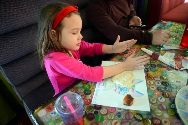
The FDA also warned that pizza dough, “play clay” made from dough, or “flour crafts” that kids sometimes play with, can also lead to food poisoning. Even if they don’t eat it, the E. coli can get onto their hands, which usually end up in their mouth before they get washed.
In light of these cookie dough poisonings, manufacturers have started to use only pasteurized eggs and, more recently, heat-treated flour to destroy the bacterial culprits. However, they still caution that consumers cook the product properly to be on the safe side.

What about those of us who enjoy our desserts within a dessert, namely cookie dough ice cream? You can breathe easy - cookie dough ice cream is okay to eat because it is typically made with heat-treated flour and pasteurized eggs.
To put things in perspective, E. coli contaminated spinach sickened nearly 200 people and killed 3 of them in 2006. The point is not to eat more cookie dough instead of spinach (sorry)…but to handle and prepare ALL food properly no matter what it is.

Putting it all together, if you are making cookie dough or cake batter from scratch, odds are the flour you're using has not been heat-treated to kill bacteria, so there is a chance it could be contaminated. Even if it is not under the recall, the flour should be treated as you would any other raw food. If you must play cookie dough roulette, some companies are stepping up to the (kitchen) plate and making some that lacks eggs and uses heat-treated flour.

Contributed by:  Bill Sullivan


Thorpe, C. (2004). Shiga Toxin--Producing Escherichia coli Infection Clinical Infectious Diseases, 38 (9), 1298-1303 DOI: 10.1086/383473

Obrig, T. (2010). Escherichia coli Shiga Toxin Mechanisms of Action in Renal Disease Toxins, 2 (12), 2769-2794 DOI: 10.3390/toxins2122769

Tuesday, June 21, 2016

Osteoporosis: The Dying Osteocyte

Bone is a highly dynamic tissue. Every year approximately 10% of an individual’s skeleton is resorbed and new bone is formed, which means that every 10 years your bones are made of entirely new material. We call this process “bone remodeling”. As we age, the balance between bone resorption and bone formation changes, leading to relatively more bone being removed and less bone being formed – this is called osteopenia, which refers to low bone mass, and is a normal consequence of aging. When the balance tilts excessively toward the loss of bone, we refer to it as osteoporosis.

Shuler F. ORTHOPEDICS. 2012    
Osteoporosis is a disease characterized by bone fragility and an increased incidence of broken bones, which results when an individual’s bones become thinner and more brittle.  Osteoporosis has long been thought to mainly affect elderly women; however, with the increasing use of prescription medicines such as glucocorticoids, and the large number of people leading unhealthy lifestyles, the incidence of osteoporosis is predicted to significantly increase in the future. In 2002, approximately 43 million people had either osteoporosis or osteopenia, and in 2020 this number is predicted to grow to nearly 61 million people.

Alterations in bone remodeling – the coupled action of bone resorption and bone formation – that lead to osteoporosis are a result of changes in the activities of the cells that carry out these processes. Bones are made up of three main types of cells:  osteoblasts, osteoclasts, and osteocytes. Osteoblasts are responsible for forming new bone, while osteoclasts eat away (resorb) the old or damaged bone. Osteocytes are osteoblasts that become entombed within the newly formed bone matrix, and they are the most abundant cell type accounting for nearly 90% of the cells. Osteocytes are the main regulators of the osteoblasts and osteoclasts. Osteocytes are among the main producers of the cytokine receptor activator of nuclear factor kappa-B ligand (RANKL) and the decoy cytokine receptor osteoprotegerin (OPG). The ratio of RANKL:OPG controls osteoclast formation because OPG is able to bind to RANKL and prevent its binding to the RANK receptor. On the osteoclast precursor surface, the cytokine RANKL binds to the RANK receptor and activates osteoclast differentiation and function of the osteoclasts, leading to increased bone resorption. The osteocytes embedded in the bone connect to each other through outgrowths called cannaliculi, creating networks within the bone that allow for the bone to sense mechanical stimuli and transmit signals between the cells.

www.medscape.com
Connexin (Cx) 43, a key protein involved in the formation of gap junctions, which are intercellular channels between the cells that allow for cell-to-cell communication. As an individual ages, the levels of Cx43 decrease and the number of dead osteocytes increases. Animal models with an osteocyte-specific deletion of Cx43 display increased osteocyte cell death, empty lacunae (the spaces in the bone cortex normally occupied by living osteocytes), and an increased number of osteoclasts along the bone surface. Experiments studying MLO-Y4 osteocytic cells lacking Cx43 also found an increase in cell death. Transfection of the Cx43 back into these osteocytic cells was sufficient to prevent this increase in cell death observed in this cell line. Osteocytes lacking Cx43 undergo a specific form of programmed cell death called apoptosis. The process of apoptosis is initiated through the action of multiple caspase proteins, including caspase-3. This increase in osteocyte apoptosis leads to the release of specific molecules and signals, which are involved in communicating with the osteoblasts and osteoclasts.
www.medicographia.com. 2012
To study the effects that osteocyte apoptosis has on osteoclast recruitment, we collected the conditioning media (the media containing growth factors that is added to cells) from Cx43-silenced and control MLO-Y4 cells that were either untreated or treated with DEVD, a caspase-3 inhibitor. This conditioning media was then used to treat non-adherent bone marrow cells that were treated with m-CSF (macrophage colony stimulating factor) and RANKL to induce osteoclast differentiation. This study found that blocking osteocyte apoptosis reduced the levels of soluble RANKL and prevented the increase in osteoclast recruitment and activity associated with osteocyte cell death.

The overall findings from this study suggest that Cx43 is required to maintain osteocyte viability and show that the increased osteoclast activity observed in Cx43 silenced osteocytes is a result of the increased osteocyte apoptosis. These findings provide a potential way in which osteocyte apoptosis could be targeted to prevent bone fragility in individuals with low bone mass.

Currently, the majority of osteoporosis drugs on the market work to maintain bone mass through inhibiting the activity of the bone resorbing osteoclasts. While these drugs are effective at preventing further bone loss, they do not reverse the bone loss that has already occurred before treatment has begun. This is because bone formation and resorption are coupled in bone remodeling*, so inhibition of resorption also decreases the amount of formation. The findings from this study provide evidence that specifically targeting osteocytes could allow for a therapeutic method to prevent bone loss and maintain bone mass through mechanisms that do not involve completely inhibiting the activity of osteoclasts.

* The uncoupled action of bone formation and bone resorption is referred to as “bone modeling”, which is one of the ways that bones can change their shape as we grow during childhood and adolescence.

Contributed by:  Hannah Davis

References:


Shuler, F., Conjeski, J., Kendall, D., & Salava, J. (2012). Understanding the Burden of Osteoporosis and Use of the World Health Organization FRAX Orthopedics, 35 (9), 798-805 DOI: 10.3928/01477447-20120822-12

Tuesday, June 14, 2016

Our Bodies Were Not Built To Last

As summer kicks in to high gear, the weather is heating up, MLB pennant races are heating up, barbecue grills are heating up, and the music is reckless and hot. Last summer witnessed the 50th anniversary of the Grateful Dead with an epic reunion and the 20th anniversary of the passing of Jerry Garcia. Yet, unbelievably, the music never stopped and the long strange trip continues. The Dead have been resurrected once again, with Dead and Company rocking a six-week US tour. With my kids finally accepting their own fate as the next generation of Deadheads, the revival of the music that makes so many of us feel alive has gotten me thinking about heritability and what information (biological or existential) is critically important to pass on to the next generation. And further, what is the meaning of life? (Full disclosure: this could simply be how I am dealing with my own process of aging, so please humor me for a moment…)
 
Here’s a sobering thought: the fundamental meaning of life is to get your genetic material into the next generation. There is no deeper meaning from a biological perspective than to maximize reproductive fitness. Different species take different approaches to spreading their seed, so-to-speak. On one end of the spectrum, some animals like sockeye salmon and mosquitos, which live in relatively unstable environments where the probability of survival is relatively low, focus on producing large numbers of offspring before they die, hoping that these offspring will reach sexual maturity and also reproduce. This is a good reproductive strategy in such environments because reproductive fitness is defined not by one’s ability to pass on their own genes, but by their offspring’s ability to pass on their genes.
 
Contrast this with the approach at the other end of the spectrum. Some animals, like orcas and primates live in much more stable environments, in population densities that are near carrying capacity for the environment (the highest density that can be supported by the resources available). These species produce many fewer offspring, but because there is a high probability of survival for offspring and parents alike, the parents invest a considerable amount of energy into raising their offspring (I know this is true because my kids exhaust me! Please help…)
 
In the case of primates, in particular, we know a great deal about reproduction, life history, and parental investment: primates produce few offspring, invest considerable energy into offspring care, and they generally have lengthy lives relative to other species in the same habitats. Compound this with advances in modern medicine, and human primates are enjoying lifespans that increase with successive generations. We live longer than our grandparents’ generation, and so on and so forth. And while this is fantastic news because we get to enjoy our children and grandchildren for longer than at any other time during human history, this does not come without a significant price: our skeletons break down in ways that other primates’ do not. Simply put, our bodies were not built to last.
 
Humans are unique among primates in that we walk around on two legs. In fact, the evolution of our bipedal locomotion predated the evolution of our large brains by several million years. And our unique mode of locomotion combined with our ever-lengthening lifespans has resulted in several musculoskeletal problems that we develop as we age. Before we get to that somber topic, it is useful to review some of the anatomical adaptations that allow us to walk around bipedally.


1. Forward position of foramen magnum. The foramen magnum – or the opening in the skull through which the brain stem/spinal cord exit – is more anterior positioned in humans compared to other primates and mammals, which places the vertebral column directly underneath the skull as opposed to behind it as in quadrupedal animals.

2. S-shaped curvature of vertebral column. By moving the vertebral column directly underneath the skull, humans require an S-shaped spinal curvature (with cervical and lumbar lordoses and a thoracic kyphosis) in order to balance the head and torso over the pelvis.

3. Broad pelvis with laterally-flared iliac blades. The iliac blades of the human bony pelvis – the parts that stick out to the side – are rotated laterally and flare outward from the midline of the body. This positions the lesser gluteal musculature (gluteus medius and gluteus minimus) lateral to the hip joint, enabling these muscles to function as abductors of the thigh at the hip joint, and prevent excessive pelvic tilt to the unsupported side during the stance phase of bipedal gait. In nonhuman great apes, this musculature is positioned posteriorly and acts synergistically with the gluteus maximus to extend the thigh, not abduct it.

4. Oversized hip and knee joints. Joint loading in response to bipedal locomotion, as well as that reflective of body mass, is borne entirely through the joint surfaces of the lower limb in humans. Therefore, we evolved expanded articular surfaces compared to our great ape relatives, which reduces shear stress in the articular cartilage. The femoral condyles and tibial condyles of the human knee are also significantly flatter in lateral profile than in nonhuman apes, which further reduces shear stress in articular cartilage. Because articular cartilage is avascular and cannot actively repair itself, reducing the shear stress borne by the cartilage also reduces the incidence of damage.

Carrying angle of the femur
5. Carrying angle of femoral shaft. The shaft of the human femur (thigh bone) is oriented obliquely relative to the femoral condyles (the part of the femur that sits on the tibia, or leg bone, to form the knee joint). This angled shaft places the knee joint directly under the center of mass. In quadrupedal animals (including our great ape relatives), the femoral shaft is more vertically oriented.

6. Adducted hallux. The human hallux – or big toe – is in line with all other digits of the foot, enabling an efficient toe off in an anterior direction in bipedal gait. In nonhuman primates, the hallux is abducted, which enables these animals to grasp with their feet in a fashion similar to manual grasping.


7. Sesamoids in tendons of flexor hallucis brevis. Sesamoid bones are bones that develop in the tendons of muscles, and the best example is the patella, or knee cap. In humans, sesamoids also develop in the tendon of flexor hallucis brevis, a muscle in the sole of the foot that flexes the big toe. These sesamoids create a tunnel through which courses the tendon of flexor hallucis longus (another big toe flexor muscle). This tunnel allows flexor hallucis longus to remain free to contract and flex big toe when all of the body weight is placed on head of the 1st metatarsal, such as when pushing off during walking.


These seven adaptations to bipedal locomotion are present in the earliest members of the fossil genus Australopithecus (and some earlier ones too), even before brains evolved to be bigger. So one can make the argument that bipedal locomotion is the hallmark of human evolution, with the evolution of big brains being a secondary adaptation that may or may not be related to the evolution of our unique locmotor mode.

Numerous hypotheses exist as to why we evolved this weird form of walking. Walking around bipedally is energetically efficient; it requires only approximately 1 calorie/min to walk. Was this the advantage it proffered over quadrupedal locomotion? Or perhaps we became bipedal in order to free our hands up to carry provisions back to our mates. Or maybe it was a way of reducing heat stress by reducing the surface area where sunrays hit directly while increasing the amount of surface area exposed to wind? Could it have evolved in order for us to see over tall grasses in the savannah? Or to increase feeding efficiency and resource exploitation? Or perhaps it was so we could posture for mates… All of these are plausible hypotheses, and there are plenty of scientific arguments in favor or one or more of these. But the fact remains, it doesn’t really matter why we evolved bipedal locomotion. Any way you slice it, we evolved it. And now we’re saddled with the baggage of our ancestors: bodies adapted to bipedal locomotion take a severe beating. Again, our bodies, especially our skeletons, were not built to last.

Vertebral compression fracture
Bone is approximately 60% mineral (calcium and phosphate) and the other 40% is collagen and other proteins. We reach our peak bone mass at about 30 years of age, which means that the most responsive time for us to build bone mass is while we are young and growing. With age, everyone loses bone mass and density; we call this osteopenia, and it is normal. But when we lose an abnormal amount of bone mass, we can this osteoporosis. Osteoporosis is common, with about 54 million Americans suffering from this disease, and often results in bone fracture. The most prevalent osteoporotic fractures are vertebral compression fractures, where the loss of bone in the vertebral column results in fracture of the body of the vertebra itself. Humans and the other great apes have an equivalent amount of bone mineral and equal bone densities, but human vertebral bodies are enlarged to absorb more compressive shock during bipedal locomotion. Therefore, they have thinner walls of the vertebral body, which are at risk of collapsing with reduced bone mass and/or density, resulting in compression fracture. These types of fractures do not occur in nonhuman apes because they have thicker walls of their vertebrae than do humans, and because their spines are parallel to the ground, not perpendicular, so there is no axial compression of the vertebral column during locomotion.


Another result of repetitive compression loading of the spine that only humans suffer is degenerative disc disease. Intervertebral discs between each of the vertebrae of the spine are comprised of two tissue types: a central, jelly-like nucleus surrounded by a strong, fibrous ring that contains the nucleus. After repeated compression, the fibrous ring of the intervertebral disc can break down, leading to a posterior bulge that impinges upon peripheral nerves that exit the spinal cord (which runs through the canal in the posterior aspect of the vertebral column). Ergo, pinched nerves. These degenerated discs do not occur with high frequency in nonhuman apes, again as a result of their quadrupedal (and less destructive) mode of locomotion.

Degenerative disc disease
One final example of how our long lifespans are not in accord with the “lifespan” of our skeleton is degenerative joint disease and osteoarthritis. The ends of bones with joint spaces are covered in a thin layer of hyaline (articular) cartilage. Hyaline cartilage is avascular, meaning that it does not have its own blood supply, and it cannot actively repair itself if damaged. In order to protect against damage, joint surfaces (of the knee at least) become flatter as body size increases during growth. This is because as body mass increases, so does the transarticular load transmitted through joint surfaces (and hyaline cartilage covering them). This has the effect of reducing shear stresses that are experienced by the cartilage and limiting the capacity to damage the cartilage by growth alone. When the hyaline cartilage breaks down, the result is damage to the bone, pain, and joint swelling – osteoarthritis. The incidence of osteoarthritis in nonhuman great apes is dramatically lower than in humans, and is attributed to a combination of shorter lifespans in the wild and the lack of a destructive, bipedal mode of locomotion.


Given all of the ways that our skeletons break down during life, it is truly quite remarkable that 60-70 year old musicians such as the surviving members of the Grateful Dead are still able to shake it, shake it in the summer of 2016. So we should embrace it while we can keep on dancing, keeping in mind that while every cloud has a silver lining; in this case, every silver lining does have a touch of grey.


Contributed by: Jason Organ, PhD


Jungers, W. (1988). Relative joint size and hominoid locomotor adaptations with implications for the evolution of hominid bipedalism Journal of Human Evolution, 17 (1-2), 247-265 DOI: 10.1016/0047-2484(88)90056-5


Jurmain R (2000). Degenerative joint disease in African great apes: an evolutionary perspective. Journal of human evolution, 39 (2), 185-203 PMID: 10968928


Latimer B (2005). The perils of being bipedal. Annals of biomedical engineering, 33 (1), 3-6 PMID: 15709701


Russo, G., & Kirk, E. (2013). Foramen magnum position in bipedal mammals Journal of Human Evolution, 65 (5), 656-670 DOI: 10.1016/j.jhevol.2013.07.007


Ward, C. (2002). Interpreting the posture and locomotion ofAustralopithecus afarensis: Where do we stand? American Journal of Physical Anthropology, 119 (S35), 185-215 DOI: 10.1002/ajpa.10185